BioUnfold #27 — Organ-on-Chip: Biology as a Living Interface
Restoring the interfaces experiments removed
Most discovery systems are built on a practical assumption: that biological effects can be separated and measured independently.
Assays are designed so that changes in a readout can be attributed to a defined perturbation. Cells are plated individually, pathways are tested outside tissue context, and targets are evaluated without circulation, barriers, or neighboring organs. Upstream experimentation works because signals can be assigned locally and interpreted without interference from surrounding biology. This strategy is extremely effective. Separability makes experiments faster, clearer, and scalable, and most questions in early discovery can be answered this way.
Organ-on-chip systems allow selected interfaces between tissues to be restored while keeping experiments observable and interpretable.
Biology is organized before it is molecular
Cells do not behave the same way when they are isolated from their environment. They respond differently when exposed to gradients, when exchanging signals across barriers, or when positioned next to another tissue rather than plated on plastic. These differences are not experimental noise. Transport between compartments, exposure timing, and spatial proximity often determine whether a mechanism becomes visible at all.
Standard cell assays remove this structure to make measurements easier to interpret. Organ-on-chip systems restore part of that structure without immediately losing control of the signal. They make it possible to study relationships between tissues rather than only perturbations within them.
A spectrum rather than a device
Organ-on-chip is not a single technology. It describes a way of structuring experiments so that selected interfaces between tissues remain present during measurement.
At one end of the spectrum, patterned co-culture systems place multiple cell types in defined locations within standard plates while remaining compatible with high-throughput workflows. At the other end, microfluidic platforms connect engineered organoids through perfused channels that simulate circulation between tissues, sometimes turning those channels themselves into functional biological compartments. Between these extremes lies a continuum of systems that progressively restore spatial organization without fully restoring organism-level complexity.
Organ-on-chip systems therefore do not increase complexity uniformly. They restore only the structure required to observe a specific interface.
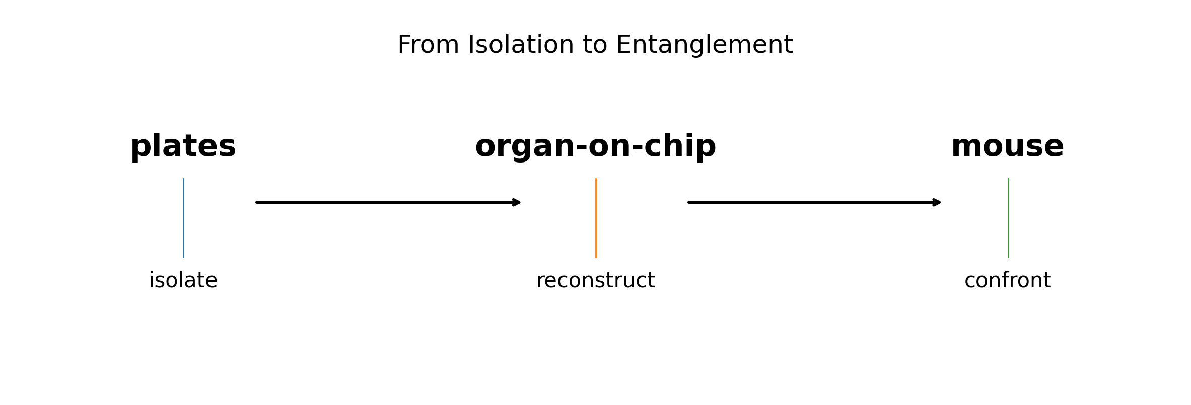
Between plates and mice
Plate-based assays isolate mechanisms efficiently but remove physiological structure. Mouse models restore systemic biology but also restore variability, adaptation, and pharmacology across the whole organism. Organ-on-chip systems occupy the space between them by reintroducing selected biological interactions while keeping experiments observable and interpretable.
Unlike mouse studies, they are usually built from human cells. This makes them particularly valuable when transport, exposure, or tissue-specific signaling differs between species, as is often the case in neuroscience, metabolism, and inflammatory disease.
Separability remains the dominant strategy in discovery because most effects can be measured independently. Organ-on-chip systems become useful when the question itself depends on interactions between compartments rather than activity within a single component. In these situations, isolating elements removes the phenomenon being studied.
This is common across therapeutic areas. In oncology, tumors evolve through interactions with immune cells, vasculature, and surrounding stroma, and therapeutic response increasingly depends on modifying those relationships rather than targeting tumor cells alone. In neuroscience, the blood–brain barrier determines whether therapies reach neural tissue at all, often deciding whether a mechanism remains actionable. In metabolic disease, communication between liver, circulation, gut, and adipose tissue shapes disease progression, and conditions such as ischemia or steatohepatitis emerge from disrupted oxygen delivery, exposure gradients, and inter-organ signaling rather than from single pathways.
Organ-on-chip systems make these interfaces experimentally accessible without immediately moving to whole-organism studies.
Why adoption remains uneven
Organ-on-chip systems improve decisions only if they are introduced early enough to change them. When introduced late, they often contradict assumptions already embedded in targets, assays, and program design, and timelines rarely allow those assumptions to be revised.
They also do not replace a single incumbent model. Instead, they overlap with plate assays used for screening, barrier assays used for transport, and animal models used for systemic validation. At the same time, they produce data that is harder to capture and interpret than standard plate readouts. Signals emerge across compartments and time rather than in a single measurement channel, and extracting meaning often requires imaging, temporal sampling, or multi-modal analysis pipelines that are not yet standard across discovery organizations. Their value therefore depends less on capability than on timing and integration within the experimental workflow.
Biology as a living interface
Most discovery systems work by separating biological effects so they can be measured independently. This strategy is extremely powerful, but it also removes many of the interfaces through which biological systems operate.
Organ-on-chip experiments do not replace reductionist biology. They allow parts of that structure to be rebuilt in controlled form. By selectively restoring interfaces between tissues, they make it possible to test how much of a biological effect depends on the surrounding organization rather than on the isolated components themselves.
They do not restore full organisms. They reconstruct just enough structure to reveal where experimental simplification begins to fail.
Biology is not only molecular. It is relational.